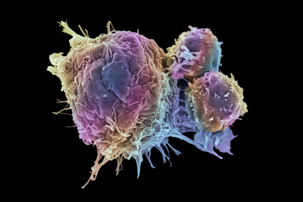
Cancer growth cells are responsible for partition tenaciously, shaping strong tumors, or flooding the blood with irregular cells. Cell division is a typical procedure utilized by the body for development and fix. A parent cell partitions to frame two little daughter cells, and these small daughter cells are being used to manufacture new tissue or to supplant cells that have kicked the bucket as a result of maturing or harm. Solid cells quit separating when there is never again a requirement for more little daughter cells. However, malignant growth cells keep on creating duplicates. They are likewise ready to spread, starting with one piece of the body then onto the next in a procedure known as metastasis.
CLASSIFICATION
There are various classes of malignant growth cells, characterized by the cell type from which they originate.
- Carcinoma, most malignant growth cells are epithelial in the starting point in the membranous tissues that line the surfaces of the body.
- Leukemia starts in the tissues liable for creating fresh recruits cells, most usually in the bone marrow.
- Lymphoma and myeloma got from cells of the invulnerable framework.
- Sarcoma begins in connective tissue, including fat, muscle, and bone. The focal sensory system got from cells of the body and spinal line.
- Mesothelioma begins in the mesothelium, the covering of body pits.
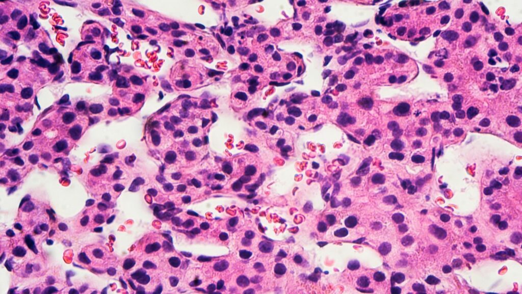
HISTOLOGY
Cancer cells have to distinguish histological features visible under the microscope. The nucleus is often large and irregular, and the cytoplasm may also display abnormalities.
The shape, size, protein composition, and texture of the nucleus are often altered in malignant cells. The core may acquire grooves, folds, or indentations, chromatin may aggregate or disperse, and the nucleolus can become enlarged. In healthy cells, the nucleus is often round or ellipsoid in shape, but in cancer cells, the outline is often irregular. Different combinations of abnormalities are characteristic of different cancer types, to the extent that nuclear appearance can use as a marker in cancer diagnostics and staging.
CAUSES
Disease cells are generated when the qualities answerable for directing cell division got harmed. Carcinogenesis is brought about by transformation and epimutation of the hereditary material of typical cells, which agitates the ordinary harmony among multiplication and cell demise. This outcome in uncontrolled cell division and the advancement of those cells by common choice in the body. The unbridled and regularly fast expansion of cells can prompt considerate or dangerous tumors (malignant growth). Favorable tumors don’t spread to different pieces of the body or attack different tissues. Life-threatening tumors can attack various organs, spread to inaccessible areas (metastasis), and become perilous.
More than one change is vital for carcinogenesis. A progression of a few changes to specific classes of qualities is generally required before a normal cell will change into a malignant growth cell.
Harm to DNA can be brought about by presentation to radiation, synthetic substances, and other ecological sources; however, changes likewise usually aggregate after some time through uncorrected mistakes in DNA interpretation, making age another hazard factor. Oncoviruses can cause particular kinds of malignant growth, and hereditary qualities are additionally known to play a role.
Undeveloped cell research proposes that an abundance of SP2 protein may transform undifferentiated cells into disease cells. However, an absence of specific co-invigorated particles that guide in the manner antigens respond with lymphocytes can impede the healthy executioner cells’ capacity, eventually prompting malignancy.
Pathology
Cells assuming jobs in the invulnerable framework, for example, T-cells, are thought to utilize a double receptor framework when they decide if to slaughter debilitated or harmed human cells. Off chance when a battery pressurized, transforming into tumors, or tainted, atoms including MIC-An and MIC-B are created so they can join to the outside of the cell. These work to enable macrophages to recognize and murder malignancy cells.
Discovery
Early proof of human disease can decipher from Egyptian papers (1538 BCE) and preserved remains. In 2016, a 1.7 multi-year old osteosarcoma was accounted for by Edward John Odes (a doctoral understudy in Anatomical Sciences from Witwatersrand Medical School, South Africa) and associates, speaking to the most seasoned recorded dangerous hominin cancer.
The comprehension of malignancy was altogether best in class during the Renaissance time frame and into the Age of Discovery. Sir Rudolf Virchow, a German researcher, and lawmaker contemplated minuscule pathology and connected his perceptions to an ailment. He depicted as “the author of cell pathology.” In 1845, Virchow and John Hughes Bennett freely watched fine increments in white platelets in patients. Virchow accurately distinguished the condition like a blood malady and named it leukämie in 1847 (later anglicized to leukemia). In 1857, he was the first to depict a kind of tumor called chordoma that began from the clivus (at the base of the skull).
Telomerase
Disease cells have one of a kind highlights that make them “godlike,” as indicated by sure analysts. The compound telomerase is utilized to expand the malignancy cell’s life expectancy. While the telomeres of most cells abbreviate after every division, in the long run making the cell kick the bucket, telomerase broadens the cell’s telomeres. A significant explanation that malignant growth cells can amass after some time, forming tumors.
Researchers found that an atom outside of a tumor seems to advance medication opposition—by changing over the tumor cells again into a foundational microorganism like state.
At the point when the tumor cells started to display sedate opposition, the cells were all the while changing into an immature microorganism like state, which made them impenetrable to the medications. It created the impression that the treatment itself was driving this change by initiating a particular atomic pathway. Fortunately, a few existing drugs, for example, Bortezomib, for instance, can assault this pathway and invert the cell change, consequently ‘re-sharpening’ the tumor to treatment.
Treatment
In February 2019, restorative researchers reported that iridium appended to egg whites, making a photosensitized particle, can infiltrate malignancy cells and, after being illuminated with light (a procedure called photodynamic treatment), annihilate the disease cells.
Link to similar posts; The Radium girls, a tale os oblivious poisoning

Fatima Zahra is a student of Quaid-e-Azam University Islamabad doing her BS Biochemistry. she is interested in reading suspense stories, creative writing, and also fond of cooking.
