23.8 C
Pakistan
Tuesday, July 8, 2025
Home Blog Page 21

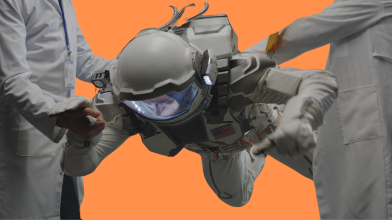
How NASA keeps astronauts healthy

The International Space Station (ISS) is a marvel of modern technology and human ingenuity, orbiting 408 kilometers above Earth at a speed of 28,000 kilometers per hour. The ISS is an incredibly complex machine, composed of several modules, laboratories, and living quarters, all designed to sustain human life in the harsh conditions of space. As such, the health of the astronauts on board is of paramount importance to NASA, and the agency has implemented a number of measures to ensure their well-being.

One of the primary ways in which NASA controls astronaut health on the ISS is through the selection and training process. NASA carefully screens all potential astronauts for medical issues that could be exacerbated by spaceflights, such as heart problems, vision impairments, and psychological disorders. Those who pass the screening undergo rigorous training that includes physical fitness, emergency procedures, and medical procedures. Astronauts also receive extensive medical training to allow them to diagnose and treat their own medical conditions while on board the ISS.

Once an astronaut is selected and trained, NASA closely monitors their health throughout their mission. Each astronaut undergoes regular medical evaluations to assess their physical and mental well-being, including blood tests, vision tests, and psychological assessments. Any medical issues that arise are promptly addressed, either by the astronaut themselves or by medical professionals on the ground. NASA has a dedicated team of flight surgeons who provide medical support to astronauts both on the ground and in space, and who are available 24/7 to address any medical emergencies that may arise. Astronauts can call their flight surgeon, who might direct them to a drug in the medical kit, or give other suggestions. This technique is known as TeleMedicine.

In addition to monitoring astronaut health, NASA also takes measures to prevent the spread of disease on board the ISS. Since the ISS is a closed environment, astronauts are at risk of developing illnesses that could spread rapidly throughout the crew. To minimize this risk, NASA employs a number of strategies to prevent disease outbreaks.

In addition to monitoring astronaut health, NASA also takes measures to prevent the spread of disease on board the ISS.

One key strategy is quarantine. Before launching to the ISS, astronauts spend several weeks in quarantine to ensure that they are not carrying any infectious diseases. During this time, they are isolated from other people and closely monitored for any signs of illness. This helps to prevent the introduction of pathogens into the closed environment of the ISS.

Once on board the ISS, astronauts follow strict hygiene protocols, including regular hand washing and the use of disinfectants, to prevent the spread of germs. They also take care to isolate any crew members who become ill to prevent the spread of disease.

NASA is conducting ongoing research on the microbiome of the ISS to better understand the complex microbial communities that exist within it. This research has revealed that the microbiome of the ISS is quite different from that of Earth, and that certain bacteria and fungi are more prevalent in space. By studying these differences, NASA hopes to better understand how microorganisms affect human health in space, and how to prevent the spread of disease in closed environments.

Finally, NASA is constantly innovating new technologies and techniques to improve astronaut health on the ISS. For example, NASA is currently developing a new medical diagnostic device that can quickly and accurately diagnose a wide range of medical conditions in space, allowing astronauts to receive timely treatment without having to rely on medical professionals on the ground. One device of this type is the HemoCue, small blood-sampling device that counts white blood cells within minutes from a single fingerstick sample. NASA is also investigating new ways to produce food in space, which could help to improve the nutritional quality of astronaut diets and reduce the risk of food-borne illness.

In conclusion, the health and well-being of astronauts on the ISS is of paramount importance to NASA. Through a combination of careful selection and training, ongoing monitoring and medical support, disease prevention strategies, microbiome research, and technological innovation, NASA is constantly striving to ensure the safety and health of astronauts in space. As space exploration continues to advance, NASA will undoubtedly continue to develop new strategies and technologies to improve astronaut health and safety.

References:

NASA, Clinical Infectious Diseases, Harvard Business Review

Also read: HISTORY OF SPACE EXPLORATION

An Ode to Parasites: How infections made us who we are today…

Let’s get this straight right away: Bacteria and viruses, specifically the infectious and harmful type have practically propelled us through millennia, molding our genomes into their current state, allowing us to become the apex species that we now are. In a nutshell, we owe our uniqueness, our sexual mode of reproduction and even an arguably premature death to these foes.

You might be wondering, what has prompted the author to accredit the despised, disease-causing, sickness-inducing microbes with such a monumental achievement. What, you may think, could be the role of these little troublemakers, when the entire body, day in and night out is pouring out an endless stream of defense mechanisms against them? At the first glance, it doesn’t make sense that organisms that our body fights tooth and nail to keep out, could be the very architects of our genome.

But here’s the plot-twist in the saga: the pathogens have never intended anything beneficial for us, indeed they’ve been trying their very best to bring us down. But this has backfired for them every single time. Let’s travel back to the very beginning of life.

How it started?

Nearly 700 million years ago, our first multicellular grandparents crawled out of the murky waters of Earth. However, upon emerging they found their surroundings wholly infested with single-celled and even colonizing micro-organisms. Those micro-organisms had spent the last 3 billion years competing for resources and niches (habitats) to fill.

It did not take them long to infiltrate the tissue of the multicellular newbies. They walked into the bodies of the multicellular organisms and began making adjustments to their new home. They poked and prodded, ate from the tissue and secreted toxins. In essence, they became parasitic and caused sickness.

All this in the name of selfishly adapting to their new hosts. This is where a bit of Darwinian Magic or Divine Will allowed mutations in the genomes of the multicellular organisms. The same mutating force, which had led them to the threshold of multicellularity, was not about to abandon them at this stage. The events of genetic variations between successive generations ultimately allowed some (or one) of the mutants to adapt to the parasitic problem of the microbes and survive the first onslaught.

The wheel gets rolling

As the multicellular organisms saw the first amendment/addition of resistance to their genome in the aftermath of their first battle with the microbes, the tone for the majority of evolution was set. From there on, our multicellular grandparents fought countless such wars with the microbes, and similar to sustaining post-war scars, they collected genome sequences or “genes” resistant to a myriad of diseases.

Their genomes grew in size, complexity, and sophistication. So much so that millions of years later, we have an enormous genetic corpus of additions from each of those encounters. For instance, our pathogen-recognition genes or HLA (Human Leukocyte Antigen) have no less than 12,000 variants1.

To put that in perspective, that is almost a hundred times more varied than any other part of our genome. This is an anomaly in stark contrast with the rule of thumb for evolution, which tends to simplify the genome of any unnecessary and burdensome variations. To say that 12,000 is a cumbersome quantity of variations to retain is an understatement.

Multicellular organisms
Our pathogen-recognition genes or HLA (Human Leukocyte Antigen) have no less than 12,000 variants

According to the modern understanding of our genetic pedigree, the only reason we have not gotten rid of most of these variants, many of which correspond to resistance against ancient and long-forgotten diseases, is that each microbe associated with the variants keeps returning, stronger each time, having evolved in the span of days or even hours. It’s like we keep a specialized diary of each version of each infectious microbe, that is handed down the generations, and is updated with each new encounter. The shocking part is that some of the genes we carry around today are 30 million years old2.

Is a “Thank You” in order?

Even more astoundingly, these pathogens caused the refinement of the human race. According to the University of California Bio-molecularity expert, Ajit Varki, 2 million years ago, a malaria-like pathogen attacked the hominid tribe (made up of Australopithecus, Homo habilis, – erectus, and sapiens). It targeted all organisms which produced certain Sialic acids3.

It manipulated the acids to gain access to the interior of the host cells, wherein it fulfilled its parasitic tendencies, causing the cell and then the host to collapse. In simple terms, due to its potent virulence and lethal effects, it caused the eventual extinction of all the members of the hominid tribe except a few deficient in sialic acid production. These few would go on to become the forefathers of Homo sapiens or modern humans.

Enter, Sexual reproduction!

It was to avoid such disasters of complete extinction on the part of host species that only after one billion years of exposure to pathogenic aggression, sexual reproduction was taken up by them; simply to create as many genetically unique variants of themselves in hopes of avoiding death at the hands of a single-minded pathogen. Before, these organisms had to rely completely on genetic mutations between asexually produced generations. However, as the word “mutation” suggests, there wasn’t much thought going into the process. More often than not, the mutations would produce variations that worsened the genome, downright reducing the organism’s chances of survival.

As a counterpoise, sexual reproduction evolved which allowed the organisms to make sober decisions about mixing their genome with another superior and preferred genome. Even today, if a woman inexplicably finds herself attracted to the broad chins or deep-set eyes, or even the body odor4 of a potential mate, it is entirely thanks to the internal biological mechanism picking up hormonal or pheromonal signs of health and virility, since austere facial features signal high testosterone levels while contrasting body odor is a sign that their offspring will be immunologically well-endowed.

All shall taste death

Looking closely at the inner structure of the DNA, we are surprised to find “Suicide Genes” that are responsible for automatically turning off the cell machinery at a certain time frame. To a rational mind, the fact that our body refuses immortality when it is entirely within reach, is strange to say the least. Why our cells simply stop dividing at a certain age is one of the greatest secrets of human biology.

Indeed, organisms around us like trees, have not opted out of the possibility of immortality. They continue to live as long as they are sheltered from physical and chemical trauma, and certainly only grow stronger as they age. Why we become weaker with each passing year while trees grow mightier is also answered by the role of pathogens in our long history with them.

Looking closely at the inner structure of the DNA, we are surprised to find “Suicide Genes” that are responsible for automatically turning off the cell machinery at a certain time frame.

The drawback of being an immortal species, as Sonia Shah puts it, is quickly expanding to occupy its limits in an ecosystem. Under normal circumstances, such an outcome is quite favorable, but in the chance of a natural disaster or more appropriately a “pandemic” from parasitic microbes means that the entire species stands at a risk of being wiped off the face of Earth. Similar to how a sparsely spaced forest has a greater chance of surviving a fire than a thick and dense one.

The Red Queen

This theory of evolution of sex and suicide genes5, is jointly called as the “Red Queen Hypothesis6, a grim reference to Lewis Carroll’s Alice in Wonderland, where despite running for a while, Alice can’t seem to move away from where she is. To this, the Red Queen of Hearts tells Alice she must run twice as fast to get anywhere!

Similarly, the relationship between pathogens and hosts is one of a constant cycle of adaptation, evolution and resistance, with both parties having ran significantly at the end of the day, with no real change in the balance of power. If the pathogen mutates to become more viral or infectious, after some initial success, it begins to falter as the host adapts and becomes resistant to its mechanisms.

It is the consequences of this cat-and-mouse strategy of evolution that our bodies spend enormous amounts of energy patrolling our tissue against pathogens, with thousands of white blood cells, extremely acidic and basic mediums and layered membranes. Essentially, making us who we are as organisms, in regards of genetic complexity, sophistication of defenses and a general psychological bias against anything remotely sickness-inducing.

Outlook

Even today, as surgeons rush to transplant organs and suppress the immune reactions of host patients, as women subconsciously pair with men that boast opposite immune system configurations from their own, and as new pathogens jump the boundary from the wild into humans in novel cases of zoonosis, the genomic grounds are seeing imperceptible but tectonic shifts in their layout. As new genes of resistance and immunity are being added and the arsenal of protective measures grows, so does the human race evolve into its next era.

References

  • 1,2 Shah, Sonia. “Pandemic: tracking contagions, from cholera to Ebola and beyond
  • 3 Ajit Varki, “Human Specific Changes in Siglec Genes”
  • 4 Meyer and Thomson, “How selection shapes variation of the human major histocompatibility complex”
  • 5 Mitteldorf, Josh; Pepper, John. “Senescence as an adaptation to limit the spread of disease
  • 6 Hamilton, William. “Sexual reproduction as an adaptation to resist parasites

Also Read: ROLE OF GENETICS IN INFECTIOUS DISEASES SPREAD, GENETICAL EVOLUTION

Contagion: A fictional version of a real-time pandemic

Contagion is a 2011 thriller about the fictional MEV-1 pandemic, the piece that everyone rewatched in 2020. Primarily because of how the story was jaw-droppingly similar to the conditions across the globe back in the Coronavirus pandemic. 

Contagion has gone viral again, its theme of the rapid sweeping of a novel virus called MEV-1. The movie shows the rapid spread of the deadly virus that kills 26 million people worldwide. The virus spreads through coughing and sneezing, just like Covid-19.

Several other pandemic movies have recently been seen rising to the top again, but none has received as much renewed success as Contagion. This documentary cum movie released in 2011 and made by director Steven Soderbergh stars Matt Damon, Kate Winslet, Gwyneth Paltrow, and many others. It is an anxiety-inducing biological disaster horror with terrifying scientific plausibility. 

The film starts with patient zero, Paltrow, as Beth Imhoff, on day two of the outbreak.

The film starts with patient zero, Paltrow, as Beth Imhoff, on day two of the outbreak. Imhoff in the beginning, looks rough, sweating, coughing, and eating peanuts from the airport bar (not a very good idea). In the sequence, Soderberg uses come clever camera work lingering on close-ups of inanimate objects, the peanuts, Beth’s card, and the touchscreen. Later in the movie, we would find out the scientific term for these i.e., “fomites”- shared surfaces where viral infections can spread from one person to another.

These fomites are shark fin in the water, the invisible threat that the movie returns to, repeatedly. Real-life experts have compared the pandemic in this movie to the current Covid-19 situation and they grade the movie high for focusing on fomites. And in their review, they also point out that the turnaround time would need to be longer more than a few days for the virus to shed from Beth’s respiratory tract or saliva to be spread to anything she’s touching.

The fictional disease in Contagion is called MEV-1. Burns and Soderberg conceived it while researching the H1N1 Flu pandemic in 2009, the 2003 SARS outbreak, and many other similar diseases. They consulted with researchers like Larry Brilliant, known for his work in eradicating smallpox, Ian Lipkin, a WHO specialist, and author Laurie Garrett.

These are the people who have dedicated their lives to studying real outbreak scenarios and conceiving hypothetical nightmare situations like one presented in this film. Beth dies two days later. MEV-1 moves super-fast, and that is the big part of the danger. Infected characters do not have much time after exposure to react or educate themselves, do self-quarantine, etc., before they accidentally pass it on to someone else. By comparison, Covid-19 seems to have a more extended incubation period reported as 14 days by WHO.

Another big difference appears to be the mortality rate. MEV has the mortality rate of between 20-30%, and by the end of the movie, it ends up killing twenty-six million people worldwide, which of course, is insanely high but not as high as the 50 million people who died in the 1918 Spanish Flu! The covid-19 mortality rate is still being determined, but as of March 3rd, it is mentioned to be 3.4% of reported cases. And the number will keep on fluctuating with time. 

The disaster response in Contagion is roughly accurate to the one these days. In the movie, WHO deploys an epidemiologist Dr. Orantes to Hong Kong to try to identify its source; meanwhile, the CDC deploys CIS officer, Dr. Mears, to the cluster region in Minneapolis and to try to contain it. While all this happens, more terms are thrown to help the audience understand this disease.

Dr. Mears explains how one sick person is likely to infect others, R0 (reproductive rate of the virus). In the movie, this value is two and later jumps to four. The value for covid-19 is estimated at approximately 2.5, and again, that doesn’t make it deadlier; it is just a lot more infectious than the seasonal flu that comes every year. 

The movie also introduces the CDC scientist Dr. Hextall who breaks down the MEV in the form of colors. She explains that the sequence of the virus contains both bat and pig sequences. The final scene of the film reveals the near accidental day one event. It shows Beth’s mining company, which deforests a jungle and displaces some bats that eat bananas.

A banana chunk drops in a pigpen, a pig eats a banana and ends up in the kitchen. And from one thing to the next, the chef touches pig’s mouth, doesn’t wash his hands, affects Beth, and Beth becomes a deadly carrier.

The science in the movie is mostly accurate, which is something to be appreciated as most of the movies don’t really consider that. Another notable research by Burns is on ecotones. Humans invade remote areas and expose the food chain to microbes, and their immune systems aren’t biologically prepared. Covid-19 stems from a family of beta Coronaviruses that all have their origins in bats.

Returning to the story, Dr. Hextall knows that for developing a vaccine, it’ll take months and months to reach human trials and FDA approval. This pushed her to take an enormous risk, and she ends up testing a vaccine strain on herself. Luckily it worked! This particular moment is an homage to physician Barry Marshall who inoculated himself with Helicobacter pylori to prove it was the cause of gastric ulcers. He was awarded the Nobel Prize in 2005 for his heroic contribution. 

The genius of Contagion is the way it tackles the crisis with a large team of people working in various fields. Disease outbreaks make a community of specialists work together while the rest of us are obliged to listen to them and to abstain from freaking out too much. 

The smart characters in the film make a lot of great heroic decisions. However, there are also occasional bad decisions, and the least heroic among them is Allen Crum Weedy, a social media influencer. He first breaks the footage of an early infected person and uses his followers to spread conspiracy theories and pedals snake oil cures like the homeopathic treatment called Forsythia. Misinformation spread by people like Crum Weedy is the true Contagion of Contagion. Indeed, nothing spreads like fear. 

Also, read: Your Guide to the Movies with a Pandemic

Role of genetics in infectious diseases spread, genetical evolution

0

Conventionally, infectious diseases were thought to be an environmental or extrinsic factor that spread through pathogenic microbes like bacteria, viruses, fungi, and various parasites. For this reason, killing infectious pathogens or avoiding the point of contact was thought to be the primary goal in preventing the spread of infections, also known as communicable diseases.

However, with the advent of ‘the genetic theory of infectious diseases in the 1920s, scientists and the world community understood better that various other factors are also responsible for the spread, as well as the aggressiveness of infectious diseases and the most critical of them all is the role of genetics. 

Genetics, particularly genetic evolution, has played a pivotal role in infectious disease spread and its morbidity and mortality rates. Therefore, to control infectious diseases, it is imperative to understand the role of genetics in both the pathogen and host in affecting infection pathogenicity, transmission and spread.

Why do some people fight infections better than others?

Host genetic predisposition and asymptomatic nature of infectious diseases

Infections do not always show the same symptoms in everyone. Recently observed during Corona pandemic, many cases went undiagnosed as individuals showed no apparent infection symptoms (the asymptomatic cases) while others suffered normal to aggressive symptoms (the symptomatic cases). 

This difference arises from the individual ‘Genetic Makeup,” which differs across a population, making the host immune stronger to fight against infection and therefore show no symptoms, or have weak immunity that leads to a slower response against the infection and therefore exhibits the disease symptoms. This ‘Genetic Predisposition’ leading to a weak response is primarily the result of mutations, making such individuals more vulnerable to infection and its spread.

To better illustrate the idea, let’s look into an example of AIDs by HIV infection, the susceptibility of which increases in individuals having mutated CCR5 (Chemokine Receptor 5) gene. CCR5 is a crucial component of the host immune response signaling expressed on lymphocytes, a key player in the immune system. In addition, the HLA (Human Leukocyte Antigen), a gene complex, is a significant factor in host immune response and susceptibility to various infections.

Genetical evolution in infectious pathogens

Pathogens constantly strive to adapt to the environment, undergoing constant genetic evolution, making them more virulent, and aiding their survival.

“Not only the host, but genetic variations in pathogens also affect the disease outcome”

The main culprit behind infections getting severe, resistant, and transmissible is the genetic evolution of pathogens causing them. This genetic evolution in microbial genomes often results in new strains that may resist previous treatments. This happened when MRSA (Methicillin Resistant Staphylococcus Aureus) strain, causing most skin infections, emerged out of S. aureus (Staphylococcus aureus).

Genetic evolution may also result in novel disease emergence, as it happened in the case of coronaviruses acquiring repeated mutational and recombinational variations, resulting in a novel variant, SARS-COV-2, responsible for the Covid-19 pandemic.

How do infectious pathogens acquire genetic variability to evolve?

Different genetic processes make pathogens evolve and thrive in the environment. Some of these are: “Mutations, Recombination, Horizontal Gene Transfer, and Selection Pressure”.

Genetic mutations 

Pathogens primarily depend on their power to rapidly mutate their genetic makeup, allowing them to survive in the environment. These mutations may be spontaneous (happen during DNA replication) or induced by the environment. Environmental factors are particularly important to understand as they may help us control pathogen evolution.

The environment-induced mutations mostly happen when drugs, chemicals, or radiations are employed with the intention of wiping out pathogens. However, it results in new and resistant strains. An exciting example is the influenza (flu) virus, which undergoes rapid mutations inside the host, changing its surface proteins to evade host immune recognition. 

Genetic recombination

This method of achieving genetic variability is quite interesting. Genetic recombination involves the exchange of genetic material between 2 different strains of the same pathogen, particularly when they come close together while infecting the same host. This lets new pathogenic strains emerge, showing improved virulence and survival features. Genetic recombination of influenza viral strains between domestic livestock and humans may result in severe consequences such as a pandemic.

Horizontal gene transfer

This process usually occurs in bacteria and plays a critical role in bacterial evolution. Horizontal gene transfer is an inverse process of vertical gene transfer. Whereas vertical gene transfer allows the transfer of genetic material and traits from parents to offspring (same species), horizontal gene transfer lets two distant species (not related by descent) share genetic material.

Horizontal gene transfer may be accomplished through any of the following three methods: 

Conjugation, where two cells directly exchange DNA by fusing at conjugative pili or through plasmids, e.g., Agrobacterium tumefaciens and E. Coli undergoes conjugation.

Transduction, where viruses called bacteriophage infect different bacteria, injecting the DNA from one to another e.g., E. coli bacteria has been transduced through lambda phage virus to allow it to use biotin and galactose. 

Transformation, which allows the integration of foreign DNA, is taken up by a competent cell. These processes have resulted in various antibiotic, drug, and pesticide-resistant bacteria e.g., Streptococcus pneumonia.

Selection pressure 

This allows infectious pathogens to thrive by adapting to their environment. Selection pressure is the external force that makes a pathogen evolve to suit its survival better. These pressures may include host immunity, transmission mode, host population pressure, or any danger in the environment, such as drugs, vaccines, and antibiotics used to stop pathogenic growth.

This has resulted in bacteria that have adapted to thrive in the presence of multiple antibiotics and viruses with more substantial virulence (the ability to cause disease). An interesting example of adaptation to population and immune pressure is the evolution of the influenza A virus into the H1N1 subtype, which caused a severe pandemic in 1918, leading to a high mortality rate.

Genetical evolution and the spread of infectious diseases

A solid and direct relationship exists between genetic evolution and the broader spread of infections. Infection transmission and spread greatly depend on the host’s genetic susceptibility and the microbe’s genetic and pathogenic factors.

Hosts with weaker immune are more susceptible to infection, allowing pathogens to survive and transfer to the other hosts e.g., CCR5 (Chemokine Receptor 5) mutated hosts are more susceptible to AIDs. Another relevant example is the mutated human DARC (Duffy Antigen Receptor of Chemokine) gene which allows malarial transmission.

On the other hand, genetic factors of the microbe make them more pathogenic and virulent. Let’s look into the example of the influenza virus that undergoes mutations to change its surface proteins and evade host immunity. This lets the virus survive, multiply in huge numbers, and quickly spread out to the other hosts. Another example is the genetic evolution of HCV (hepatitis C virus), which lets it transmit through multiple routes to infect a larger population.

In the case of COVID-19, the genetic change in viral surface receptor ACE2 (Angiotensin Converting Enzyme 2), allowed it to transmit and enter the host cells efficiently.
In the case of COVID-19, the genetic change in viral surface receptor ACE2 (Angiotensin Converting Enzyme 2), allowed it to transmit and enter the host cells efficiently. Photo WHO

In the case of COVID-19, the genetic change in viral surface receptor ACE2 (Angiotensin Converting Enzyme 2), allowed it to transmit and enter the host cells efficiently. In addition, many pathogens spread among hosts by genetic changes in genes responsible for virulence and transmission, such as toxins and adhesins e.g., S. aureus (Staphylococcus aureus) spreads to the host by using its antibiotic resistance ability provided by the SCCmec (staphylococcal cassette chromosome mec).

What next? Genetic-associated preventive measures!

Our understanding of host and pathogen genetics has led to a safer and more effective design of preventive measures and infection treatments. We have all witnessed the consequences of drug misuse, such as antibiotics which have resulted in the emergence of more virulent and resistant bacterial strains.

Today, individuals can opt for personal genetic testing and screen out any possibility of infection susceptibility. This will help take early precautions and avoid catching an infection by undergoing timely vaccination or prophylactic treatment to stay on the safer side. 

On the other hand, genetic sequencing of the pathogen genome helps track new mutations and identify any new strain emergence. However, genetic preventions come with a price of high ethical concerns. Genetic testing of individuals may put them in an uncomfortable spot where they may face discrimination or stigmatization. In addition, gene editing and genetic therapies raise concerns for equal accessibility by all, their long-term efficacy, and safety concerns for the world’s ecosystem.

Conclusion

Host and pathogen genetic variations play a crucial role in spreading infectious diseases. Understanding the genetic evolution and genetic factors involved in infectious diseases’ spread has helped design more effective and specific preventive solutions. Advancements in genetic technologies, such as gene editing and gene therapy, offer promising prospects for controlling and eradicating infectious diseases. However, these techniques are yet costly and thus not accessible to everyone. Our aim should be eradicating this difference in the future and making genetic treatments reachable to everyone.

Also, Read: Chronic diseases: An interplay of infections and our genome

What You Eat Matters: Nutrition and Infectious Diseases

0

As we move through life, we are offered various health pointers passed through generations with no particular origin.

“An apple a day keeps the doctor away.”

“Drink at least 8 cups of water daily.”

“Eat a lot of fruits and veggies.”

“A Man is What He Eats”

However, the question remains: Why? What makes these suggestions so accurate that they’ve stood the test of time? A simple answer would be nutrition. 

Nutrition can either help build us up or breaks us down. Moreover, it is a cardinal contributor to our health as it is the fuel that gives our body the strength to recover from setbacks, fight off unwelcome interlopers, and strengthen our immune system. Hence, it can be safe to hypothesize that nutrition significantly connects to an individual’s susceptibility to infectious diseases.

This article will examine the complex relationship between nutrition and infectious diseases in South Asia, specifically Pakistan, and examine how we can use nutrition as a defense. 

The term ‘infectious diseases, according to WHO, refers to disorders that are instigated by viruses, bacteria, and parasites that can become contagious under ideal conditions (Organization). Common infectious diseases can range from influenza, dysentery, and gastroenteritis to Tuberculosis, hepatitis, and measles.

The World Health Organization (WHO) finds that such diseases are responsible for 13·7 million deaths yearly worldwide (Gray and Sharara 2022). The statistics can be staggering; 3.1 million people died from diarrheal diseases such as cholera, typhoid, and dysentery, with most of the victims being children. In addition, Tuberculosis (TB) infected 10.6 million people, malaria 247 million, hepatitis B over 2 billion, and measles more than 9 million (Foundation 2021, Iacobucci 2022, Organization 2022, Organization 2022).

Many of these deaths can be traced back to overcrowded, economically struggling South Asian countries, specifically affecting children under the age of 5 and making up half of the overall disease prevalence (Basnyat and Rajapaksa 2004, Zaidi, Awasthi, et al. 2004, Ghose Bishwajit 2014). Pakistan is one of these countries and although various efforts have been made that have increased life expectancy from 66.9 years in 2017 to 67.3 years in 2019, it is still lagging when compared to its nieghboring countries Bangladesh, China, and Sri Lanka. 

The deaths of 71,000 children every year due to pneumonia, the estimated 3.4 million people with malaria in 2022, and the 10 million people carrying hepatitis C in Pakistan demonstrate just how pressing the issue is. (Dawn 2022, Khan 2022, Organization 2022)
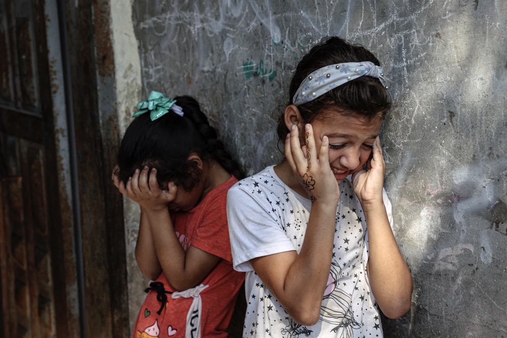
Nutrition is a cardinal contributor to our health as it is the fuel that gives our body the strength to recover from setbacks, fight off unwelcome interlopers, and strengthen our immune system.

However, recent studies have unearthed another way to combat this predicament: nutrition. With the progressive shift from agriculture to an industrial economy, there is a shift from rural to urban areas, which further populate main cities like Lahore, Islamabad, Sialkot, Faisalabad etc. This brings about a significant deterioration in the environmental conditions and lowers the standard of living and overall nutrition. Poorer nutrition leads to malnutrition, an important step that leads to an underdeveloped immune system and increased susceptibility to diseases(CHAN , Keusch 2003, Ambrus and Ambrus 2004).

This was demonstrated in a prospective, randomized nutritional intervention study that observed four groups of infants in a high-incidence malnutrition region of Lahore. The study concluded that inadequate dietary intake within the first year of life substantially increases the chances of diarrhea and malnutrition in Pakistani infants (JAVAID, HASCHKE et al. 1991).

Nutritionally compromised individuals may suffer from anorexia, decreased intestinal absorption & metabolic rate, and an overall deficiency in various multivitamins and minerals that hinder their body’s normal functioning (Farhadi and Ovchinnikov 2018). 

Improper nutrition may have lasting effects, as reported in an experimental study which concluded that Energy Restriction hindered the proper functioning of natural killer cells and increased the harshness of influenza (Ritz, Aktan et al. 2008). Not only can insufficient diet decrease immunity and susceptibility to infectious diseases, but improper diets as observed in obese individuals, can also be a contributing factor by reducing vaccine efficacy and a slowed antiviral response (Kim, Kim et al. 2011, Taylor, Cao et al. 2012, Honce and Schultz-Cherry 2019). 

Therefore, a proper diet with the right amount of nutrition can act as a game changer in the fight against infectious diseases. Not only can a nutritious diet rich in zinc, vitamins C, D, and A, along with essential omega-3 fatty acids, help buttress our immune system, but it can also ensure that our body is fit enough to tackle any illnesses (Aman and Masood 2020, Pecora, Persico et al. 2020).

An interesting point to note here is that simply increasing the amount of food you intake does not help improve your nutrition levels, but eating the right foods. We can only do this by increasing accessibility to such foods and awareness among households, especially women (Farhadi and Ovchinnikov 2018).

According to Dr Krutika, “many people don’t realize that their gut health plays a crucial role in preventing and managing infectious diseases. Consuming a diet high in fiber, fermented foods, and probiotics promotes a healthy gut microbiome, which helps strengthen the immune system and reduce the risk of infections. On the other hand, a diet high in sugar, processed foods, and unhealthy fats harms the gut microbiome and weakens the immune system, making the body more susceptible to infections.” https://www.clinicspots.com/blog/dr-krutika-nanavati-sports-dietician-nutritionist

Therefore, nutrition is essential in either exacerbating or ameliorating the progression towards or prevention of various diseases. by re-emphasizing the importance of nutrition, and how a good nutritional diet is an essential factor in preventing infectious diseases. Summarize all the points discussed in the paragraphs and suggest potential solutions for public health programs to improve dietary habits.

References

  • Organization WH. [Available from: https://www.emro.who.int/health-topics/infectious-
    diseases/index.html.
  • Gray A, Sharara F. Global and regional sepsis and infectious syndrome mortality in 2019: a
    systematic analysis. The Lancet Global Health. 2022;10:S2.
    https://www.thelancet.com/journals/langlo/article/PIIS2214-109X(22)00131-0/fulltext.
  • Iacobucci G. Measles is now “an imminent threat” globally, WHO and CDC warn. BMJ.
    2022;379:o2844. ‘doi: 10.1136/bmj.o2844. https://www.bmj.com/content/379/bmj.o2844.
  • Organization WH. Malaria-Pakistan 2022 [Available from:
    https://www.who.int/emergencies/disease-outbreak-news/item/2022-
    DON413#:~:text=Malaria%20is%20endemic%20in%20Pakistan,over%20the%20course%20of%202021.
  • Organization WH. Tuberculosis 2022 [Available from: https://www.who.int/news-room/fact-
    sheets/detail/tuberculosis.
  • Foundation HB. Hepatitis B Facts and Figures 2021 [Available from:
    https://www.hepb.org/what-is-hepatitis-b/what-is-hepb/facts-and-
    figures/#:~:text=Two%20billion%20people%20have%20been,of%20infected%20individuals%20are%20diagnosed.
  • Zaidi AKM, Awasthi S, deSilva HJ. Burden of infectious diseases in South Asia. BMJ.
    2004;328(7443):811-5. ‘doi: 10.1136/bmj.328.7443.811https://www.ncbi.nlm.nih.gov/pmc/articles/PMC383379/
  • Basnyat B, Rajapaksa LC. Cardiovascular and infectious diseases in South Asia: the double
    whammy. BMJ. 2004;328(7443):781. ‘doi:
    10.1136/bmj.328.7443.781.https://www.ncbi.nlm.nih.gov/pmc/articles/PMC383361/
  • Ghose Bishwajit SI, Sharmistha. Social Determinants of Infectious Diseases in South Asia. Ghosh
    International Scholarly Research Notices. 2014;2014(135243):10 ‘doi:
    https://doi.org/10.1155/2014/135243..
  • Khan MA. Comparative prevalence of different types of viral hepatitis in the district Dera Ismail
    Khan, Khyber Pakhtunkhwa, Pakistan. Egyptian Liver Journal. 2022;12(1):40. ‘doi: 10.1186/s43066-022-00203-1.
  • Dawn. Over 70,000 children die of pneumonia every year in Pakistan, say experts 2022
    [Available from: https://www.dawn.com/news/1686783.
  • Keusch GT. The History of Nutrition: Malnutrition, Infection and Immunity. The Journal of
    Nutrition. 2003;133(1):336S-40S. ‘doi: https://doi.org/10.1093/jn/133.1.336S.
  • Ambrus JL, Ambrus JL. Nutrition and Infectious Diseases in Developing Countries and Problems
    of Acquired Immunodeficiency Syndrome. Experimental Biology and Medicine. 2004;229(6):464-72. ‘doi:
    10.1177/153537020422900603. https://pubmed.ncbi.nlm.nih.gov/15169964/
  • CHAN HTH. Nutrition and Immunity [Available from:
    https://www.hsph.harvard.edu/nutritionsource/nutrition-and-
    immunity/#:~:text=A%20deficiency%20of%20single%20nutrients,E%20can%20alter%20immune%20responses.
  • JAVAID N, HASCHKE F, PIETSCHNIG B, SCHUSTER E, HUEMER C, SHEBAZ A, et al. Interactions
    between Infections, Malnutrition and Ironnutritional Status in Pakistani Infants A Longitudinal Study.
    Acta Paediatrica. 1991;80(s374):141-50. ‘doi: https://doi.org/10.1111/j.1651-2227.1991.tb12017.x.
  • Farhadi S, Ovchinnikov R. The relationship between nutrition and infectious diseases: A review.
    Biomedical and Biotechnology Research Journal. 2018;2(3):168-.
  • Ritz BW, Aktan I, Nogusa S, Gardner EM. Energy Restriction Impairs Natural Killer Cell Function
    and Increases the Severity of Influenza Infection in Young Adult Male C57BL/6 Mice. The Journal of
    Nutrition. 2008;138(11):2269-75. ‘doi: 10.3945/jn.108.093633.
    https://www.ncbi.nlm.nih.gov/pmc/articles/PMC2635521/
  • Taylor AK, Cao W, Vora KP, Cruz JDL, Shieh W-J, Zaki SR, et al. Protein Energy Malnutrition
    Decreases Immunity and Increases Susceptibility to Influenza Infection in Mice. The Journal of Infectious
    Diseases. 2012;207(3):501-10. ‘doi: 10.1093/infdis/jis527. https://pubmed.ncbi.nlm.nih.gov/22949306/
  • Kim Y-H, Kim J-K, Kim D-J, Nam J-H, Shim S-M, Choi Y-K, et al. Diet-Induced Obesity Dramatically
    Reduces the Efficacy of a 2009 Pandemic H1N1 Vaccine in a Mouse Model. The Journal of Infectious
    Diseases. 2011;205(2):244-51. ‘doi: 10.1093/infdis/jir731.
    https://academic.oup.com/jid/article/205/2/244/2192546
  • Honce R, Schultz-Cherry S. Impact of Obesity on Influenza A Virus Pathogenesis, Immune
    Response, and Evolution. Frontiers in Immunology. 2019;10. ‘doi: 10.3389/fimmu.2019.01071.
    https://pubmed.ncbi.nlm.nih.gov/31134099/
  • Aman F, Masood S. How Nutrition can help to fight against COVID-19 Pandemic: COVID-19 and
    Diet. Pakistan Journal of Medical Sciences. 2020;36(COVID19-S4). ‘doi: 10.12669/pjms.36.COVID19-
    S4.2776. https://pubmed.ncbi.nlm.nih.gov/32582329/
  • Pecora F, Persico F, Argentiero A, Neglia C, Esposito S. The Role of Micronutrients in Support of
    the Immune Response against Viral Infections. Nutrients. 2020;12(10):3198.10.3390/nu12103198

Also, Read: Dr. Shagufta on how nutrition can help to combat Coronavirus

The consequences of long-COVID and post-COVID

0

COVID-19, also known as Novel Coronavirus is a highly contagious disease of the respiratory system caused by the SARS-CoV-2 virus. First identified in Wuhan, China in late 2019, this virus rapidly spread across the globe resulting in mortality rate on a massive scale. It was declared a pandemic by World Health Organization (WHO) in March 2020.

The symptoms of COVID-19 range from mild to severe cases, commonly starts with the onset of high grade fever, followed by shortness of breath, sore throat, loss of taste and smell, extreme body aches, fatigue, chest congestion and cough.

The world has suffered a significant blow due to COVID-19 including the impacts of severity level on global health, social interaction, and economies. COVID-19 led to a surge in social distancing which confined everyone in their domestic zones, shifting the whole manual system to online and remote work disrupting supply chains, in-land and international travel, and distancing.

Long COVID-19 or Post-COVID

Long COVID is also known as Post-Acute Sequelae of SARS-CoV-2 infection (PASC), a condition characterized by people in which they continue to experience prolonged symptoms even after the virus is no longer detectable in their bodies.

This condition affects anyone regardless of age, severity of prior COVID infection or vaccine administration. Its symptoms develop usually after the initial infection which might last for two months or over.

Symptoms of long-COVID

The symptomatology of infection is similar to the original infection but from case to case the severity varies. It is notable that over 200 different symptoms have been reported that affected the overall functioning of the body.

The symptoms of Long-COVID are not only related to the respiratory tract but it has symptoms associated with the nervous system and other vital organs. It is more of a syndrome based on its diversified, new, returning, or ongoing symptoms of multiple organs which last for four weeks or even more after the initial COVID infection. In some reported cases post-COVID lasted months or years and caused disabilities.

Some most common symptoms of post-COVID-19 are:

  • High-grade, low grade or continuous fever
  • Respiratory congestion
  • Fatigue 
  • Recurrent coughing
  • Headache and dizziness
  • Sleep problems
  • Neurological symptoms including difficulty thinking, memory loss and lack of concentration
  • Depression or anxiety
  • Tickling , pins and needles feeling
  • Loss of taste and smell
  • Continuous pain in muscles and joints
  • Cardiac symptoms including chest pain, heaviness, rapid pounding of heartbeat
  • Digestive symptoms including stomach pain, acid reflux and diarrhea
  • Blood clots causing pulmonary embolism
  • Onset of allergies related to respiratory tract and nasal passages
  • Disturbance in menstrual cycle
  • Multi organ impairment or permanent failure including kidneys, liver, pancreas, bones and skin.

Numbers affected

According to the studies, almost 10-20% cases have been reported to be infected by SARS-CoV-2 might experience relapses of the disease as a long COVID across the globe. The exact number of living cases for this form is uncertain as many cases do not get proper documentation.

Impacts of long COVID on different organs
Impact of long COVID on different organs. Source: Nature

According to the recent statistical evidences, at least 65 million people around the world are suffering from long COVID, based on a conservative estimated incidence of 10% of the infected individuals and there are more than 651 million documented COVID-19 cases worldwide; this number is likely much higher due to many undocumented cases. The incidence is estimated at 10–30% of non-hospitalized cases, 50–70% of hospitalized cases and 10–12% of vaccinated cases.

Protection from long-COVID

Protection from this disease means to shield yourself and others from COVID-19 infection. World Health Organization (WHO) recommends a number of simple but proven measures to get rid of this illness.

  • Get regular vaccination and boosters of COVID-19
  • Wear masks
  • Wash or sanitize hands regularly
  • Keep your premises clean and well ventilated

Recovery

Recovery for this condition varies from case to case. Some symptoms can improve quickly and others take quite longer to get recovered. The anomalous behavior of Coronavirus suggests that it has no generalized affect rather from person to person this virus has had varying impacts. It devastated many healthy bodies yet embarked new life in chronic patients.

Pathophysiology

Ever since the emergence of this deadly illness, it has caused the ripples of research in the field of biomedical sciences, pathology, virology, genetics, pharmacology and drug designing. Biomedical researchers have made significant progress in identifying various pathophysiological changes and the associated risk factors in the characterization of this disease and studies are still in progress to know the root causes of the prolonged impact of this illness even after vaccination regardless of the age, health condition or gender.

The unanimous view, so far, is the auto-immunity, endothelial dysfunction, viral persistence, immune dysregulation and coagulation activation are some of the baseline pathophysiological mechanisms involved in the hauling of COVID-19.

The studies have revealed that post-COVID-19 can manifest whatever the severity of the first COVID-19 illness, the underlying pathophysiology of this long delay is still enigmatic and anomalous in general population.

References

  • https://www.cdc.gov/coronavirus/2019-ncov/long-term-
    effects/index.html#:~:text=As%20a%20result%20of%20these,have%20not%20
    had%20COVID%2D19.
  • https://duckduckgo.com/?q=COVID+19&t=chromentp&atb=v370-
    7&ia=coronavirus
  • https://www.who.int/europe/news-room/fact-sheets/item/post-covid-19-
    condition
  • https://www.mayoclinic.org/diseases-conditions/coronavirus/in-
    depth/coronavirus-long-term-effects/art-20490351
  • https://www.nhs.uk/conditions/covid-19/long-term-effects-of-covid-19-long-
    covid/
  • https://www.nature.com/articles/s41579-022-00846-2

Also Read: SCIENCE JOURNALISM NEEDS VOICES FROM SOUTH ASIA- SIRI CARPENTER

Is genetic engineering the holy grail to cure infectious diseases?

The advent of genetic engineering in recent times has shrunk the time needed to evolve any species or organism. It is an excellent amalgamation of science and the ecosystem’s natural process to meet the population’s needs. The advantages of genetic engineering are numerous, including the production of genetically modified crops and seeds, medicinal foods that can be used as vaccines, increasing the food supply, modifying organisms to exhibit traits of interest; and genetic engineering has come a long way.

The occurrence of infectious diseases has been escalating worldwide. With approximately 37.7 million people affected by HIV by 2020, 10.6 million people affected by Tuberculosis by 2021, and almost 3-5 million people/year afflicted by Influenza, it is a matter of acute concern. With the success of genetic engineering in other domains, attention is being channeled to using this technology to combat or cure infectious diseases.

“The advance of genetic engineering makes it quite conceivable that we will begin to design our evolutionary progress”- Isaac Asimov

Employing genetic engineering for the treatment of infectious diseases means introducing specific genes that discretely inhibit or block the functions of target genes of interest or impede certain gene products. The main aim of the entire process is to restrict the replication of infectious organisms (viruses, bacteria).

The success of gene therapy to treat infectious diseases depend upon several factors, such as recruiting pertinent target cell/ tissue, the efficiency of the vector to transfer the gene of interest to the targeted region, the required expression rate of the gene, and the production of sufficient gene products. The operation of genetic engineering to treat infectious diseases can be classified into three mainstream groups:

  1. Gene therapies based on nucleic acids, including DNA, RNA, and ribozymes

Antisense (nucleic acids complementary to the mRNA of specific genes) nucleic acid transcripts can be formulated to specifically target various regions of the genome of the infectious organism. The antisense nucleotides are degraded by nucleases which inhibit their translation into mRNA and thus result in no functional protein production. 

  • A benefit of antisense RNA to be used for inhibitory purposes of target genes is its lack of immunogenicity which means that they do not generate any immune reaction in the body.

Ribozymes are antisense RNA molecules that play their role in the process of gene therapy by binding to the target gene/molecule of interest, cleaving the bond at the target site, and thus preventing any replication of the division of the nucleic acid. 

  • One of the advantages of utilizing tailored RNA is that they are not used up in the process, so it can be used multiple times to achieve the desired effect. Moreover, ribozymes can be produced from small transcriptional units, and thus, they can be produced in large quantities targeting multiple genomic regions of the infectious agent via a single vector construct. Their catalytic property allows them to be excellent gene expression inhibitors at low concentrations, thus making them a significant candidate for genetic engineering.
  1. Immunotherapeutic approaches such as genetic vaccines

One of the ways to prevent the body against infectious disease is to expose the protein components of the infectious agent to the body’s immune system, thereby eliciting an immune response in the body. Such as immune response will allow the body to fight the infection and reduce the severity of the disease by allowing the immune system to combat the infection.

A prominent example is DNA-based vaccines, which are developed by transferring DNA plasmid into target cells by genetically engineering the DNA.

  1. Gene therapies based on the alteration of protein moieties, such as single-chain antibodies

There are three primary protein moieties: anti-infectious cellular proteins and trans-dominant harmful proteins.

  • Anti-infectious cellular proteins are those recruited from normal cellular genes exhibiting specific inhibitory activities. The inhibitory action of these proteins can enable their binding to the infectious agent and directly/indirectly inhibit cellular components of the infectious agent, thus inhibiting viral gene expression.
  • Trans-dominant Negative Proteins (TNPs) are mutants of the structural or regulatory proteins that demonstrate negative phenotype, a trait that can inhibit the replication of infectious agents. However, a critical issue with these moieties is that when genetically altered/engineered, they may activate the body’s immune system, resulting in self-destruction. Such an occurrence ultimately reduces the efficacy of antiviral gene therapy and defeats its purpose of curing infectious diseases.
  • Single Chain Antibodies, or intrabodies, are single-chain, intracellularly expressed antibodies. They can directly bind to the gene of interest, interact with them and inhibit gene function, thereby deranging the life cycle of the infectious agent and preventing its replication and proliferation. 

CRISPR: A revolutionary tool

Else known as Clustered Regularly Interspaced Short Palindromic Repeats, CRISPR/CAS 9 is a groundbreaking genetic editing tool that plays a significant role in infectious disease research. An array of various infectious diseases can be treated with CRISPR, including viruses, fungi, and bacteria.

diseases
  • How does CRISPR/CAS-9 work?

CRISPR/CAS works in three steps to prevent repeated viral attacks on bacteria.

  1. Adaptation

The invading virus inserts its small DNA fragments into the CRISPR sequence as new spacer molecules.

  1. CRISPR RNA production

CRISPR repeats and spaces residing in bacterial DNA undergo transcription producing a single-chain RNA molecule. CRISPR RNA cuts this newly formed RNA into shorter pieces.

  1. Targeting 

CRISPR RNA channels the molecular machinery of the bacterial host cell to destroy the viral components.

Human Immunodeficiency Virus (HIV) is an RNA virus that integrates its genetic material into host genomes. CRISPR can be used to specifically target and remove the DNA from the host cell genome. A high-resolution CRISPR screen in human CD4+ T cells was developed by Dr. Nevan Krogan’s lab in 2018 that identifies factors involved in HIV infection.

Fungal infections threaten the human population contributing to a total of 1.6 million deaths per year worldwide. CRISPR has allowed the creation of new approaches to manipulate and modify fungi genetically, the treatments for which are a challenge. For instance, CRISPR mutagenesis was developed in the fungus Aspergillus fumigatus.

CRISPR diagnostic tools for SARS-CoV-2, an RNA virus, have proven to be an efficacious treatment for the virus by yielding accurate results, producing faster results than PCR tests. Cardea Bio is a company involved in using CRISPR diagnostic elements for pathogen and infectious disease testing.

Monitoring, evaluating, and finding therapeutic approaches to combat the spread of infectious diseases is a dire need. Genetic engineering can be exploited to manipulate these infectious agents, thus preventing or reducing their replication to inhibit their spread. Though the focus is driven by integrating technology to combat infectious diseases, there is an excellent arena of untapped research.

“With genetic engineering, we will be able to increase the complexity of our DNA and improve the human race. But it will be a slow process because one will have to wait about 18 years to see the effect of changes to the genetic code”- Stephan Hawking

References:

Also, read: PAINFUL, AND POTENTIALLY DISFIGURING, CUTANEOUS LEISHMANIASIS IS A GROWING THREAT IN PAKISTAN

Painful, and potentially disfiguring, cutaneous leishmaniasis is a growing threat in Pakistan

0

The rash on his daughter’s face had turned into a large lesion, sore and extremely itchy. Gul Bibi was also running a high fever, and Abdul Wali was worried. The only skin doctor in Zhob district, north west Balochistan, diagnosed her with leishmaniasis, more commonly called “kal-dana” locally.

But in fact there’s little awareness of leishmaniasis in rural parts of Balochistan. “When Gul Bibi complained of rash and pain on her cheek, we thought it was a common sore, as skin diseases were widespread in our area after the historic flooding,” said Abdul Wali. But weeks passed, the itching grew worse, and the fever set in.

After her diagnosis, Wali brought his daughter to Benazir Shaheed Hospital Quetta due to the severe dearth of medicine and treatment facilities in remote Zhob.

He said the doctor in Quetta prescribed a course of injections labelled “Glucantime” – the commercial name for Meglumine antimoniate, the first-choice drug against all forms of leishmaniasis.

The cost for the treatment would run to more than 20,000 Pakistani rupees (USD 74), Wali said.

“I somehow managed expenses, but hospital authorities later informed me that they had run out of Glucantime stock. The injection wasn’t available in the open market either,” Wali recalled.

Abdul Wali is not the only worried parent to find himself with a clinical diagnosis for his daughter's illness, but without reliable access to treatment.
Abdul Wali is not the only worried parent to find himself with a clinical diagnosis for his daughter’s illness, but without reliable access to treatment.

Sandfly scourge

A parasitic infection spread by the sandfly, cutaneous leishmaniasis is spreading fast in Balochistan, with the Federal Ministry of Health reporting a spike in cases in the province in the first quarter of 2022. The majority of people affected were women and children.

Cutaneous leishmaniasis (CL) is one of three forms of the disease. Unlike visceral leishmaniasis, also called “kala-azar”, the cutaneous form is not typically fatal – but can be scarring and disabling.

In CL, the site of the sandfly bite soon develops a papule or nodule, which may eventually develop into large plaques or ulcerating lesions, depending on the particular species of the parasite, and the host’s immune response.

According to Dr Zeeshan Haider, Associate Professor of Microbiology and Biomedical Sciences at the University of Lahore, these lesions may be self-healing. However, this can take several months to years – with a typical recovery time of two to eight months. Left untreated, these so-called “Oriental sores” can develop into chronic disfiguring lesions or swelling of the affected parts, inhibiting mobility.

According to Dr. Zeeshan Haider, these lesions may be self-healing; however, this can take several months to years, depending on the species. Leishmaniasis major lesions usually heal within two to eight months. If left untreated, they can develop into chronic disfiguring lesions or swelling of the affected parts, further causing disability and disfigurement.

The sandflies mainly bite on exposed body parts, like the face, legs, feet, and hands. More often than not, these lesions leave scars leading to psycho-social problems, stigmatisation, and discrimination in families and communities, especially for women, said Dr. Zeeshan.

A growing problem

According to WHO data, 186,703 leishmaniasis cases were reported across Pakistan between 2014 and 2018. Balochistan province, where more than 60,000 cases were reported in that time period, appeared to be the epicentre already at the time.

Balochistan registered 21,000 in the 12 months between January 2021 and 2022, showing an accelerating spread of the disease. That uptick was concentrated in rural areas.

Dr Mubashir Khan, a specialist at Children’s Hospital Quetta, said that a major factor behind the recent increase in leishmaniasis cases in both Sindh and Balochistan provinces has been migration from Afghanistan, with most cases reported in the border lands.

Other significant factors are climate change, urbanisation, poor hygiene, and sanitation conditions in rural areas. Sandflies thrive among mud and unplastered houses, especially where livestock are abundant.

A deadly relation

That’s a potentially deadly concern. Sandflies also spread CL’s more dangerous cousin, visceral leishmaniasis, which, if untreated, is fatal in almost every case. WHO estimates 50,000-90,000 new cases of VL a year, concentrated in Brazil, east Africa and India.

Dr Shahzeen Saifullah, a skin disease expert in Karachi who handled what may have been the first reported case of successfully-treated visceral leishmaniasis from Gilgit Baltistan, told VaccinesWork just how grave VL can be – even handled in a hospital setting.

A sick five-year-old child from the city of Swat was brought to him at the Civil Hospital Karachi. According to Dr Saifullah, the boy suffered from high grade fever and had several lesions on his nose and face. The boy was weak, suffered chills and rigors, his abdomen distended. He bled from his rectum. It took five weeks of in-patient treatment before the child stabilised, found his appetite again, and began to slowly return to health.

The disease, Dr Saifullah suggests, is very often missed or misdiagnosed.

Prevention and cure

Abdul Wali is not the only worried parent to find himself with a clinical diagnosis for his daughter’s illness, but without reliable access to treatment.

Low stocks of effective medicines at regional health centres mean that thousands of CL patients from rural areas make the journey to the Medicins Sans Frontiers (MSF) leishmaniasis centres at Quetta and Kuchlak in Balochistan, and Peshawar in Khyber Pakhtunkhwa.

According to Dr. Zeeshan Haider, Glucantime is an effective drug for treating cutaneous leishmaniasis in central Iran. In fact, cutaneous leishmaniasis heals spontaneously, and to prevent the acquisition of resistance, the indications for treatment in each region should be defined carefully.

The Drug Regulatory Authority Pakistan (DRAP) has issued an alert that certain agencies or pharmacies used to sell non-regulated/ false medicine (Glucantime) with the same name in Quetta and the rest of Pakistan.

The leading company claimed to be manufacturing this has confirmed that they do not manufacture this product and neither they sub-contract the manufacturing, nor distribute these products anywhere in the world, said Dr. Zeeshan.

According to Shahzad Badar, field communication officer at MSF Pakistan, MSF has been treating leishmaniasis at Kuchlak in Balochistan since 2008.

“In Balochistan, three well-established cutaneous leishmaniasis centres are providing free treatment,” Badar said. “In 2022, nearly 5,974 patients received treatment from [Khyber Pakhtunkhwa] and Balochistan clinics.”

Badar explained that prevention and control of the sandflies requires effective integrated vector control management, which is highly complex. “Therefore, our primary focus is on early diagnosis and effective treatment to reduce the prevalence and, thus, the transmission.”

MSF is training up local corps of lady health workers (LHWs) to spot and spread awareness of this growing public health problem. These lady health workers visit villages to make people aware of the severity of the disease and explain preventative measures. Those include the use of chemically-treated bed nets, sealing cracks in houses, placing traps in windows, regularly removing garbage and spraying insecticides.

“We are arranging training sessions for Ministry of Health paramedics for the diagnosis and cutaneous leishmaniasis case management. To increase access to prompt diagnosis and effective treatment, we will enhance this programme to Bannu and other areas of KPK in the coming months,” added Badar. Like Balochistan’s, Khyber Pakhtunkhwa’s CL caseload has been mounting since 2018.

New strategies for the treatment

According to Dr. Zeeshan Haider, unfortunately, no vaccine against is currently available worldwide against Leishmaniasis for humans. But certain vaccines are at hand for Leishmaniosis in Dogs.

A couple of scientists are working on developing new technologies, therapies, or drug regimes. Like Nanotechnology, the use of Nanoparticles against these resistant Leishmania strains. But this is at a very nascent stage and currently invitro studies are going on, and it may have side effects.

  • Moreover, MSF has started a clinical trial in which we are studying the effectiveness of three other modes of treatment. According to Shahzad Badar, these treatments are:
  • An oral drug, miltefosine, which patients can take at home.
  • Thermotherapy, the heat delivered on the site of the CL lesions via radio frequency. It is a procedure that only requires one session.
  • The combination of these treatment modalities with a shorter course of miltefosine.

A few parts of this story have earlier been published at Gavi, The Vaccine AllainceAlliance. We republished it with the author’s permission.

Also, read: Cardiovascular diseases are the number one cause of deaths worldwide

Successful cure for HIV with Stem Cell Transplantation

Human Immunodeficiency Virus, HIV is the deadliest virus that directly attacks the immune system leaving it unable to fight off illness. Its infection progresses through stages starting with flu-like symptoms followed by an asymptomatic stage with low virus levels, which can last for years. Later persistently swollen lymph nodes and other symptoms develop, leading to severe immunodeficiency, life-threatening infection, AIDS, and Acquired immune deficiency syndrome. The decline in the immune system makes the person more prone to infections and cancers. HIV can be transmitted through direct contact with body fluids, such as sharing needles, sexual transmission, and from mother to child during childbirth or breastfeeding.

History

The first case of HIV was discovered in 1980 when a cluster of patients with rare types of pneumonia was reported among gay men in Los Angeles. Since then, more than 84.2 million individuals have been infected with the virus resulting in 40 million deaths worldwide. The development of treatment for HIV has been an ongoing process for many decades. Antiretroviral treatment is the commonly given therapy for HIV/AIDS.  But this treatment only suppresses the replication of the virus, thus improving the immune system. There was no cure for the complete remission of HIV, until the discovery of Stem cell transplantation.

Breakthrough in HIV Treatment 

Scientists found stem cell transplantation can treat various types of cancers. Recently they discovered that it could be used to cure AIDS. Stem cells are specialized cells that can transform into different types of cells.

Stem cells are like superheroes, capable of wielding the power of regeneration and healing in our body, just like the Avengers saving the world from destruction.

Stem cell transplantation is considered the most promising treatment for HIV. One of the significant benefits of stem cell transplantation for HIV treatment is that it offers a potential long-term cure rather than just managing the virus with lifelong antiretroviral therapy. Additionally, stem cell transplant has the ability to reverse the damage occurred by HIV to the immune system, which can potentially lead to complete restoration of the immune system. 

Demystifying the Mystery

HIV attacks a genus of immune cells called T-cells (a type of white blood cell) responsible for fighting against bacterial and viral infections by recognizing and attacking them. In Stem cell transplant, the T cells of infected patients are replaced with the involves replace with the healthy donor who is immune to HIV infection. The immunity comes from the rare mutation which develops resistance against HIV. When these new T cells are replaced with the infected T cells in the body of the patient. They start to attack the virus and prevent it from replication. Eventually, the virus becomes cleared from the body of patients, and these patients are considered cured of HIV.

Stem cell transplant is still a relatively new experimental approach for HIV treatment;

Successful Treatment of HIV 

The first person cured of HIV by stem cell transplantation was Timothy Ray Brown, also known as the “Berlin patient”.  He had both HIV and leukemia. His doctors decided to give him a stem cell transplant from a donor who is immune to HIV, hoping it would cure his leukemia and HIV. After the transplant, his leukemia went into remission, and his HIV was wholly cured.

He remained HIV-free for more than a decade. After that, another patient was cured of HIV by a stem cell transplant HIV-resistant mutation. He received stem cells from a donor having the same HIV-resistant mutation. Several clinical trials are currently underway to investigate the efficacy of treatment.

Conclusion

While stem cell transplantation is a promising treatment for HIV, it has its own complications. The procedure is expensive and invasive and involves the risk of complications, such as secondary infections and graft versus host disease. Graft versus host disease occurs when the donor cells attack the recipient body, which can be life-threatening. Moreover, finding a suitable donor with a rare genetic mutation that makes them resistant to HIV can be difficult as it’s a rare mutation and occurs in about 1% of the population.

Stem cell transplant is still a relatively new experimental approach for HIV treatment; the ongoing research and development in this area is a promising sign for the future of HIV and potential cures. As more research and clinical trials are conducted, we may be able to see breakthroughs in advancement in the use of stem cells to treat HIV and other diseases.

REFERENCES

  • Hütter, G. “Stem Cell Transplantation in Strategies for Curing Hiv/Aids.” [In eng]. AIDS Res Ther 13, no. 1 (2016): 31. https://doi.org/10.1186/s12981-016-0114-y.
  • Justiz Vaillant, A. A., and P. G. Gulick. “Hiv Disease Current Practice.” In Statpearls. Treasure Island (FL): StatPearls PublishingCopyright © 2022, StatPearls Publishing LLC., 2022.
  • Kandula, U. R., and A. D. Wake. “Promising Stem Cell Therapy in the Management of Hiv and Aids: A Narrative Review.” [In eng]. Biologics 16 (2022): 89-105. https://doi.org/10.2147/btt.S368152.
  • Kuritzkes, Daniel R. “Hematopoietic Stem Cell Transplantation for Hiv Cure.” The Journal of Clinical Investigation 126, no. 2 (02/01/ 2016): 432-37. https://doi.org/10.1172/JCI80563.
  • Max Roser and Hannah Ritchie (2018) – “Hiv / Aids”. Published Online at Ourworldindata.Org. Retrieved From: ‘Https://Ourworldindata.Org/Hiv-Aids’

Also, read: HIV in Pakistan – An alarming Threat

Authors: Sadaf Sarfraz, Dr. Muhammad Mustafa

Chronic diseases: An interplay of infections and our genome

We all acquire infections throughout our lives, right from the neonatal period to late old age. We go through the natural history of infections that being from incubation periods to the development of immunity and resolution. While the common person is well aware of the fact that we gain resistance against infections by actually suffering from them initially, little does the common folk realize that while our bodies’ defense systems protect us in the long run, it may also prove to be the proverbial double-edged sword.

Before we delve into the perspective behind the preset set earlier, it’s pertinent to mention the burden of chronic disease in our current day and age. Chronic and especially cardiometabolic diseases (which comprise the lion’s share ) are the culmination of multiple risk factors ranging from genetic predispositions and sedentary lifestyles to stress. However, there is one binding factor behind most, if not all, chronic diseases, and that is chronic inflammation. Inflammation is an alarm state in the body, constant stress with heightened levels of markers of biological stress. What this can do is affect every organ system of the body. 

For example, inflammation sets off in the fatty tissue and pancreas, leading to diabetes mellitus, or in the vasculature of the body leading to heart disease or even strokes. Mind it; inflammation is in no way a ticket to the land of doom and gloom. Inflammation Is an intricate process set off in our bodies in the face of infections or any other insult. This is an innate response of our immune system and vital to our survival. It is this very response when continuing unabated in response to an infection, that can lead to a host of morbid conditions in the long run and even acutely in some cases.

The whole process of mounting immune responses to antigens does not happen overnight. It starts off way back when we are babies.

The beginning

When we are born, our bodies are not used to all the different types of antigens all around, and as such, we have no protection against them. As we are gradually exposed to what the world has to offer, a specific gland in the lower part of our neck called the Thymus starts producing what I would label as little ‘armies’ or ‘clones of armies’, the lymphocytes which actually will be responsible for setting up immune responses in the future against particular antigens.

Another essential process that goes on inside the gland is eliminating those ‘clones’ that target our own tissues. A process called immune tolerance to our own tissues. It is this very process when gone haywire leads to the non-exhaustive list of autoimmune diseases where the body’s own organs are targeted by the immune system.

Immune response to the invaders

As we grow older, we continuously experience infections by different pathogens, namely viruses, bacteria, fungi, and parasites. Our immune system, on being exposed to them, builds immune responses to them and, in most cases, contains them and gives our bodies arsenal in the form of alert lymphocytes and other early response cells, which will immediately target the offending agent and contain it and the next time it makes an intrusion, contain it. The immune response which is built when we are exposed to infectious agents can be experienced by us as fever, the feeling of being unwell, fluctuations in pulse rate and blood pressure, and other organ-specific symptoms according to the organism involved. 

Genes as determinants of the immune response against pathogens

As discussed previously, the body mounts responses to every kind of antigen we are exposed to since childhood, and most of the time, we are protected against those antigens throughout life. The process of recognition of antigens in organisms is driven by genes that determine the presence of specific proteins called the ‘major histocompatibility complex (MHC)’ in the immune and basically every cell of the body. These proteins are where the antigens of invading pathogens attach and the whole cascade of immune containment and inflammation begins. In humans, the genes for the synthesis of MHC are found on chromosome 6 of the human genome1.

chronic diseases
The process of recognition of antigens in organisms is driven by genes that determine the presence of specific proteins called the ‘major histocompatibility complex (MHC)’

The pathogens attach to the MHC and are then presented to specific immune cells called T-Lymphocytes. These cells are the master controllers of the whole immune response. There are countless other receptors on different cells of different body organ systems by which pathogens enter cells and cause inflammation. So, one can fathom how our immunity and reaction to pathogens is dependent upon our genome.

Pathogens causing chronic diseases: The basis of it

As elaborately detailed earlier, the body’s response to infections may eventually be the harbinger of morbidity and disabilities in the long run. Medical literature abounds with associations between pathogens and chronic illness and even cancers, though there is no definitive cause-and-effect relationship in most cases. Strong relationships exist between many organisms with cancers and serious illnesses. 

Helicobacter Pylori and its association with gastric cancers, borrelia and its association with a constellation of symptoms of Lyme’s disease, Hepatitis B and C viruses as a cause of chronic hepatitis and multi-organ involvement, and liver cancer, Epstein Bar virus and its association with Burkitt’s Lymphoma and cancers of the throat2. Besides, numerous chronic diseases have been postulated to have etiological links with infections, e.g., type 1 and 2 diabetes mellitus, rheumatoid arthritis, systemic lupus erythematosus (SLE), and chronic ischemic heart disease. This list is non-exhaustive. 

Why the above associations Have popped up over the last few decades? The answer is multifactorial firstly because we have seen a surge in the appearance and detection of zoonotic(infections transmitted from animals to humans) infections. Secondly, newer laboratory diagnostics like Polymerase chain reaction (PCR) and other advanced immunological techniques. Thirdly, Environmental factors which have gone from bad to worse in terms of high-risk behaviors, pro-inflammatory diets and mental stress all contribute in putting our immune systems into overdrive. Plus, it’s never 1+1 in disease causality. Individual genetic variations eventually determine how a particular individual will be affected acutely and in the long run by a particular pathogen.

Multiple sclerosis(MS) is one of the most prevalent neurodegenerative disorders in the world. It affects the central nervous system, i.e., the brain and spinal cord, and presents a constellation of symptoms, including motor, sensory, ocular, bladder, and bowel problems.

Disease examples

While Type 2 Diabetes Mellitus(T2DM) is a ubiquitous disease due to its solid genetic origins and environmental triggers, the etiology of Type 1 Diabetes Mellitus is a less frequent disease but with origins that are rooted in genetic inheritance only partially. Associations have been found between human enteroviruses (HEV), Rotaviruses, Cytomegalovirus (CMV), and Mumps virus, to name a few3, 4,5, which mainly target the insulin-producing Beta cells in the pancreas causing inflammation against self-tissues (autoimmunity) and, tissue destruction and eventually Diabetes Mellitus which is insulin dependent.

But on the contrary, as per the ‘ultra-clean hypothesis’, children who are less exposed to infections due to enteroviruses during early childhood are more susceptible to infections by diabetogenic viruses as they grow older. This probably relates to the paucity of protective antibodies to these viruses due to the lack of milder frequent exposure during childhood6

However, clear-cut causality has not been established either way, but what is known is that viruses attach and enter the pancreatic beta cells through cell surface receptors which are proteins7. These proteins are synthesized from genes, and inflammatory complications start here when the viruses attach. There then is local damage to the beta cells and the damage caused by the immune cells to which the viruses attach and trigger inflammatory response8 cascades, akin to wildfire, more begets more.

This illustration provided a 3D graphic representation of a spherical-shaped mumps virus particle, that was studded with glycoprotein tubercles. The studs, colorized reddish-brown, are known as F-proteins (fusion), and those colorized beige, are referred to as HN-proteins (hemagglutinin-neuraminidase).

Cardiovascular disease (CVD) is a scourge of the modern world, from genetics to hypercholesterolemia to Diabetes. Numerous factors play their part in its genesis9. However, the role of chronic inflammation in initiating or precipitating the worsening of CVD cannot be discounted10. CVD is not just a disease localized to the vasculature of the heart. It is the end process of what goes in the whole body’s vessels. Numerous pathogens have been implicated, namely Hepatitis C virus (HCV), Human immunodeficiency virus (HIV), Chlamydia pneumonia, and Herpes Simplex virus (HSV), to name a few.

 The process of the dreaded blockage of coronary or peripheral vessels starts when there is plaque formation ( accumulation of dead immune cells, platelets, and cholesterol) and rupture. The organisms mentioned earlier have been found in plaques in the vessels’ inner linings. They have been found to enter through specific adhesion molecules on the lining surface, which these pathogens induce. These molecules then facilitate the entry of pathogens, and the process of accumulation of dead immune cells, cholesterol, and platelets starts, eventually causing rupture of the plaque, which is the harbinger of ‘heart attacks11.

Multiple sclerosis(MS) is one of the most prevalent neurodegenerative disorders in the world. It affects the central nervous system, i.e., the brain and spinal cord, and presents a constellation of symptoms, including motor, sensory, ocular, bladder, and bowel problems. This disease does not have a hereditary basis though there is a genetic predisposition augmented by environmental factors and infections to manifest the disease.

Again here, like in the previous instances mentioned, associations of this disease have been found with a few infections, of which Epstein Bar virus (EBV) is worth mentioning. This virus is notorious for triggering/ causing a host of cancers like nasopharyngeal cancer and Lymphomas12. It has also been associated with autoimmune disorders. There is a pool of data from epidemiological studies, along with studies that isolate the virus from tissues, that it is implicated in causing this disease13. Though it is worth mentioning that not all EBV infections will cause disease, it is pretty ubiquitous, and most people will have antibodies to it. 

It is a particular group of people genetically predisposed to developing MS in whom this virus will cause the transformations and cascades required for the disease. The vital mechanism proposed is the entry of the virus into B-cells (mainly lymphocytes concerned with antibody production), after which it takes over the genome of the infected cells and causes dysregulated immune responses. This culminates in the production of antibodies and an immune reaction towards the myelin sheath (covering of nerves and central neural tissue) plus also against the cells which are responsible for the protection of the sheath12.

This produces the myriad of symptoms of MS, which can be progressive and relentless. Here we can see again the interplay of proteins on the surface of cells that serve as targets for the virus and genes that produce those proteins. 

Relevance of the gene-infection-inflammation-disease interplay and novelty in treatment

The concept of infections causing chronic diseases and their prevention with vaccination has been investigated for some time now. However, the results have not been encouraging. The probable reason behind this can be the multifactorial nature of many diseases plus the individual variation in susceptibility to infections. Our response to such a conundrum should be multi-tiered.

Firstly is the establishment of causality of a disease with an infection. Secondly, measures to prevent the disease in those who are susceptible. Last but not least, genetic manipulation, to be precise in our approach, which can be pursued down the line as has been done with gene editing for hereditary diseases by CRISPR. 

In the three disease examples cited, the entry portals to the pathogens after identification can be modified to prevent the entry of the pathogens. This could drastically reduce disease burden in those who are susceptible, preventing chronic morbidity and unnecessary drug treatment and improving quality of life. Gene editing by pinpointing the genes of interest and editing or deleting the concerned loci could be the future of this aspect of chronic disease prevention/treatment.

References

  • Matzaraki, V., Kumar, V., Wijmenga, C. et al. The MHC locus and genetic susceptibility to autoimmune and infectious diseases. Genome Biol 18, 76 (2017). https://doi.org/10.1186/s13059-017-1207-1
  • O’Connor SM, Taylor CE, Hughes JM. Emerging infectious determinants of chronic diseases. Emerg Infect Dis. 2006 Jul;12(7):1051-7. doi: 10.3201/eid1207.060037. PMID: 16836820; PMCID: PMC3291059.
  • Hyoty H, Taylor KW: The role of viruses in human diabetes. Diabetologia 45: 1353–1361, 2002
  • Honeyman MC, Stone NL, Harrison LC: T-cell epitopes in type 1 diabetes autoantigen tyrosine phosphatase IA-2: potential for mimicry with rotavirus and other environmental agents. Mol Med 4: 231–239, 1998
  • Pak CY, Eun HM, McArthur RG, Yoon JW: Association of cytomegalovirus infection with autoimmune type 1 diabetes. Lancet 2: 1–4, 1988
  • Viskari H, Ludvigsson J, Uibo R, Salur L, Marciulionyte D, Hermann R, Soltesz G, Fuchtenbusch M, Ziegler AG, Kondrashova A, Romanov A, Kaplan B, Laron Z, Koskela P, Vesikari T, Huhtala H, Knip M, Hyoty H: Relationship between the incidence of type 1 diabetes and maternal enterovirus antibodies: time trends and geographical variation. Diabetologia 48: 1280–1287, 2005 
  • Ylipaasto P, Klingel K, Lindberg AM, Otonkoski T, Kandolf R, Hovi T, Roivainen M: Enterovirus infection in human pancreatic islet cells, islet tropism in vivo and receptor involvement in cultured islet beta cells. Diabetologia 47: 225–239, 2004
  • .Dotta F, Censini S, van Halteren AG, Marselli L, Masini M, Dionisi S, Mosca F, Boggi U, Muda AO, Prato SD, Elliott JF, Covacci A, Rappuoli R, Roep BO, Marchetti P: Coxsackie B4 virus infection of beta cells and natural killer cell insulitis in recent-onset type 1 diabetic patients. Proc Natl Acad Sci U S A 104: 5115–5120, 2007
  • Roth GA, Mensah GA, Johnson CO, Addolorato G, Ammirati E, Baddour LM, Barengo NC, Beaton AZ, Benjamin EJ, Benziger CP, Bonny A, Brauer M, Brodmann M, Cahill TJ, Carapetis J, Catapano AL, Chugh SS, Cooper LT, Coresh J, Criqui M, DeCleene N, Eagle KA, Emmons-Bell S, Feigin VL, Fernández-Solà J, Fowkes G, Gakidou E, Grundy SM, He FJ, Howard G, Hu F, Inker L, Karthikeyan G, Kassebaum N, Koroshetz W, Lavie C, Lloyd-Jones D, Lu HS, Mirijello A, Temesgen AM, Mokdad A, Moran AE, Muntner P, Narula J, Neal B, Ntsekhe M, Moraes de Oliveira G, Otto C, Owolabi M, Pratt M, Rajagopalan S, Reitsma M, Ribeiro ALP, Rigotti N, Rodgers A, Sable C, Shakil S, Sliwa-Hahnle K, Stark B, Sundström J, Timpel P, Tleyjeh IM, Valgimigli M, Vos T, Whelton PK, Yacoub M, Zuhlke L, Murray C, Fuster V; GBD-NHLBI-JACC Global Burden of Cardiovascular Diseases Writing Group. Global Burden of Cardiovascular Diseases and Risk Factors, 1990-2019: Update From the GBD 2019 Study. J Am Coll Cardiol. 2020 Dec 22;76(25):2982-3021. doi: 10.1016/j.jacc.2020.11.010. Erratum in: J Am Coll Cardiol. 2021 Apr 20;77(15):1958-1959. PMID: 33309175; PMCID: PMC7755038.
  • Lopez-Candales A, Hernández Burgos PM, Hernandez-Suarez DF, Harris D. Linking Chronic Inflammation with Cardiovascular Disease: From Normal Aging to the Metabolic Syndrome. J Nat Sci. 2017 Apr;3(4):e341. PMID: 28670620; PMCID: PMC5488800.
  • Naga Venkata K Pothineni, Swathi Subramany, Kevin Kuriakose, Lily F Shirazi, Francesco Romeo, Prediman K Shah, Jawahar L Mehta, Infections, atherosclerosis, and coronary heart disease, European Heart Journal, Volume 38, Issue 43, 14 November 2017, Pages 3195–3201, https://doi.org/10.1093/eurheartj/ehx362
  • Soldan, S.S., Lieberman, P.M. Epstein–Barr virus and multiple sclerosis. Nat Rev Microbiol 21, 51–64 (2023). https://doi.org/10.1038/s41579-022-00770-5
  • Ascherio A, Munger KL. Epstein-barr virus infection and multiple sclerosis: a review. J Neuroimmune Pharmacol. 2010 Sep;5(3):271-7. doi: 10.1007/s11481-010-9201-3. Epub 2010 Apr 6. PMID:

Also, read: The Nabateans: A History Preserved in Rocks